Narrowing in on Neurotrophic Keratitis
Don’t fall into the trap of associating no pain with dry eye disease.
KEY TAKEAWAY
A quiet patient can still have a dangerous cornea, and a simple sensitivity check may be what prevents persistent staining from becoming a sight-threatening ulcer.
Because neurotrophic keratitis (NK) rarely announces itself with pain, we must distinguish it from dry eye disease (DED), so we can prescribe the right treatment. This column covers how we can do that.
ACQUIRE HISTORY AND ASSESS COMPLAINTS
We should ask the patient about the chronic use of preserved topical medications, especially those that contain benzalkonium chloride. Additionally, we should ask about any history of cataract, corneal, glaucoma, or retina surgeries. Finally, we should inquire about herpetic disease, diabetes, and any neuro-ophthalmic history. The reason: All these items are associated with the development of NK.
NK patients may report blur, photophobia, or irritation.

EMPLOY THE SLIT LAMP
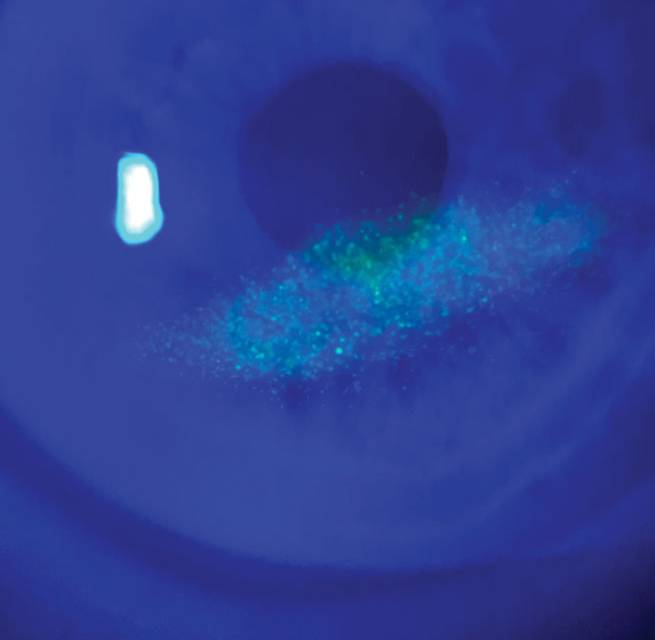
A quiet, persistent, ovaloid epithelial defect or an area of staining, especially just inferior to the pupil or near the visual axis, should raise suspicion for NK (Figure). A major red flag is if the patient doesn’t express a level of pain expected from the corneal staining or epithelial compromise present.
TEST CORNEAL SENSITIVITY
Using a cotton wisp, dental floss, or a Cochet-Bonnet esthesiometer, evaluate corneal sensitivity centrally and in all quadrants, particularly when herpetic disease, prior surgery, or neuro-ophthalmic disease is part of the patient’s history. A normal cornea should detect a light touch; a neurotrophic cornea may require a shorter, stiffer filament for a response, or it may not respond at all. A response below 6 cm should immediately shift the diagnosis from DED-induced corneal staining to impaired corneal innervation.
MANAGEMENT
This begins with reducing toxic exposure to the cornea and protecting the epithelium. To accomplish this, we can employ bandage or scleral contact lenses, an amniotic membrane, autologous serum tears, or cenegermin-bkbj (Oxervate, Dompé). Treatment depends on NK severity. Bandage or scleral lenses, an amniotic membrane, or autologous serum tears provide biologic support and growth factors to aid epithelial healing. Oxervate supplements recombinant human nerve growth factor.
BETTER SAFE THAN SORRY
A quiet patient can still have a dangerous cornea, and a simple sensitivity check may be what prevents persistent staining from becoming a sight-threatening ulcer.
FORWARD-THINKING OUTLOOK
Routine screening in high-risk patients will help ODs identify reduced corneal sensitivity and early epithelial changes before NK progresses. Do this by adding corneal sensitivity testing to ocular surface examinations, documenting staining and epithelial defects consistently, using preservative-free treatment strategies, and staying up to date as more NK treatment options emerge and evolve.
Ready to Claim Your Credits?
You have attempts to pass this post-test. Take your time and review carefully before submitting.
Good luck!