
Cataract Surgery: Identifying Postoperative Adverse Events
Know what to look for and how to intervene.
KEY TAKEAWAYS
- If the anterior chamber is formed and the Seidel leak is slow, a bandage contact lens should be placed, and the patient should be checked daily until resolution of the leak.
- Delaying treatment of endophthalmitis puts the patient at risk of permanent vision loss or loss of the eye.
- Capsular bag distension syndrome requires creating an opening in the posterior capsule using a yttrium aluminum garnet laser.
- Keep a watchful eye on patients who have a history of uveitis, vitreomacular traction, epiretinal membrane, or macular holes, as they are at an increased risk of cystoid macular edema after cataract surgery.
Although cataract extraction is one of the most common and successful ophthalmic procedures, as with any ophthalmic procedure, postoperative adverse events can occur. Because part of our collaborative care role in cataract surgery is to identify these complications and intervene, this article covers how we can accomplish both (see “What a Typical Postoperative Patient Looks Like”).
ABNORMAL IOP
We must be aware of IOP spikes and low IOP:
• IOP Spikes. IOP spikes result from retained viscoelastic, retained lens material, inflammatory debris, iris pigment release, or red blood cells that cause outflow issues in the trabecular meshwork.1 This is a common and typically temporary finding. An IOP spike of 28 mm Hg or greater occurs in 18% to 45% of postoperative patients, with most measurements returning to normal limits (10 mm Hg to 21 mm Hg) within 24 hours.2 A total of 1.3% to 10% of patients can present with IOP greater than 30 mm Hg after 24 hours.2
• Treatment. This revolves around knowing the mechanism of the IOP spike. Typically, if a postoperative patient’s IOP ranges from 25 mm Hg to 28 mm Hg, one drop of an aqueous suppressant in the clinic is instilled. Aqueous suppressants are an effective topical medication for IOP reduction due to their quick onset with direct target of the ciliary body to decrease aqueous production. If IOP ranges from 28 mm Hg to 35 mm Hg, an aqueous suppressant is prescribed for 1 week. If IOP is greater than 35 mm Hg, it is time to consider “burping” the incision in the clinic.
When burping the wound, pressure should be applied to the limbal side of the paracentesis to force aqueous out of the incision. The aqueous leaving the incision site, similar to a positive Seidel sign, should be visibly seen.
• Pro Tip. Do not burp the wound too quickly or too firmly. This is to ensure IOP has not dropped too low. Note: Patients may need to follow up before the 1-week postoperative visit for closer IOP monitoring to ensure pressure control.
• Low IOP. If a patient presents with a single-digit IOP, that may indicate a clear corneal wound leak. If a shallow anterior chamber and/or low IOP are present, Seidel sign should be tested. To do this, a fluorescein strip over the wound site is applied, and a cobalt blue filter is used to examine the area. If a clear fluid stream appears, this is a Seidel-positive sign, indicating a wound leak.
• Treatment. If the anterior chamber is formed and the Seidel leak is slow, a bandage contact lens should be placed, and the patient should be checked daily until resolution of the leak. If the Seidel leak is quick or the anterior chamber isn’t formed, send the patient to the surgeon.
• Pro Tip. These patients should be educated on the increased risk of infection with bandage contact lens placement and, therefore, the need for continued antibiotic use during wear, typically QID. If the wound continues to leak, the surgeon may recommend a suture or glue.
WHAT A TYPICAL POSTOPERATIVE PATIENT LOOKS LIKE
On postoperative day 1, we should expect blurred visual acuity (VA), corneal edema, anterior chamber cell/flare, and a centered IOL. Additionally, patients may still have a dilated iris, slightly elevated IOP, discomfort, and photopsia. The photopsia typically occurs from peripheral reflections from the IOL, which result in dark or light-curved reflections in the patient’s vision. That said, photopsia can also be a symptom of retinal detachment, so we should dilate and investigate.
At 1 week post-surgery, we should expect improved VA, IOP, and reduced inflammation/edema.
At the 1-month mark, a spectacle prescription is typically issued, and anterior chamber inflammation should be completely resolved.
ENDOPHTHALMITIS
This adverse event results from bacterial invasion into the anterior chamber, either during surgery or immediately after. Its onset is typically 3 to 7 days postoperatively, with many patients becoming symptomatic approximately 72 to 96 hours after.3 Between 40% to 80% of endophthalmitis cases are caused by cataract surgery, with 70% due to coagulase-negative Staphylococci, 10% from Staphylococcus aureus, and 9% from Streptococci.4
Symptoms are blurred and/or decreased vision, eyelid swelling, eye pain (though not always), excessive tearing or discharge, floaters, and photophobia. Clinically, endophthalmitis appears as a mild to severe anterior chamber reaction, hypopyon, fibrinous membrane, vitreous cells, retinal hemorrhage, and optic nerve head congestion.
• Treatment. This is a true ocular emergency that requires a retina consultation the same day. Delaying treatment puts the patient at risk of permanent vision loss or loss of the eye. Treatment includes a vitreous tap with cultures, intravitreal antibiotics, and possible vitrectomy, along with oral antibiotics.
• Pro Tip. Because preoperative lid disease, such as blepharitis, increases infection risk, we should identify and treat it prior to surgery.

CAPSULAR BAG DISTENSION SYNDROME (CBDS)
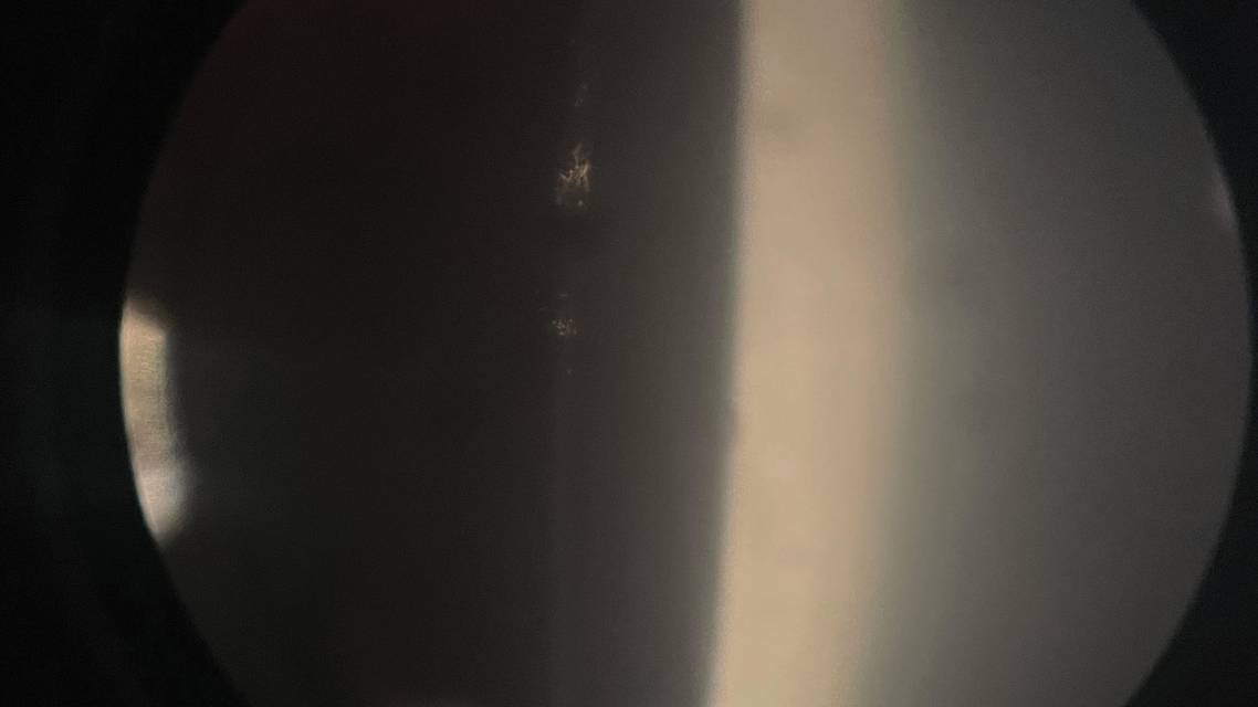
This occurs when retained viscoelastic is in the bag behind the IOL (Figure). There is a collection of retrolenticular fluid that appears clear, milky white, or turbid with expansion of the capsular bag. The patient symptoms are onset of myopia, along with gradual blurred, cloudy, dim, foggy, and/or hazy vision, reduced visual acuity, and glare that typically occurs weeks to years after cataract surgery. A positive diagnosis is a posteriorly displaced capsule that appears bowed.
• Treatment. This requires creating an opening in the posterior capsule using a yttrium aluminum garnet laser.
• Pro Tip. If the posterior capsule remains intact with clear fluid behind the IOL, this is classic CBDS.
CYSTOID MACULAR EDEMA (CME)
This may occur weeks to months after cataract extraction. The symptoms are worsening vision, persistent blurry vision, difficulty reading fine print, and mild image distortion. Clinically, CME presents as cyst-like fluid accumulation in the macula with hyporeflective intraretinal spaces. On angiography, there is a petaloid pattern of fluorescein leakage.
• Treatment. For this condition, treatment involves topical nonsteroidal antiinflammatories (typically QD to QID, depending on the medication used) and topical corticosteroids (typically QID) for a duration of 1 month.
If the CME is nonresponsive to topical therapy, an intravitreal or a sub-Tenon’s corticosteroid may be administered. Intravitreal anti-VEGF and systemic steroids and nonsteroidals may also be considered.
• Pro Tip. Keep a watchful eye on patients who have a history of uveitis, vitreomacular traction, epiretinal membrane, or macular holes, as they are at an increased risk of developing CME after they undergo cataract surgery.
STAY VIGILANT
Overall, cataract extraction is a safe and effective surgery. Outcomes are typically excellent, but complications can threaten vision if missed. It is our role to identify these risks, know the pathology, and conduct prompt management or referral as needed.
FORWARD-THINKING OUTLOOK
“Robotics, with their ultra-precise surgical movements, microscopic real-time imaging, and integrated AI” may not only improve patient safety and postoperative outcomes for cataract surgery patients, but also enhance productivity, as surgeons could potentially utilize more than one at once. This is according to a recent study in Eye.
Ready to Claim Your Credits?
You have attempts to pass this post-test. Take your time and review carefully before submitting.
Good luck!
Recommended
 Cataract/Refractive Surgery
A Review on Identifying and Explaining Cataracts
Nicholas J. Bruns, OD, FAAONicholas J. Bruns, OD, FAAO- Cataract/Refractive Surgery
Establishing a Practical Collaborative Care Protocol
Gabrielle Anderson, OD; Mitch Ibach, OD, FAAOGabrielle Anderson, OD; Mitch Ibach, OD, FAAO