Study Uncovers Novel Synaptic Intricacies Inside the Retina

A Northwestern Medicine study has uncovered novel cellular mechanisms within the retina, findings that researchers say could help advance the development of targeted therapeutics for diseases and conditions impacting vision. The findings were published in Nature Communications.
Cone synapses inside the eye’s retina help the brain process changes in light. It’s a unique synapse because it is has evolved to signal changes in light intensity, said Steven DeVries, MD, PhD, the David Shoch, MD, PhD, Professor of Ophthalmology and senior author of the study.
“Counterintuitively, cone neurotransmitter release is high in the dark and reduced by light. When the light’s brighter, the reduction is larger. When the lights are dimmer, it’s smaller; it operates differently from most synapses which use an increase in transmitter release to signal all-or-nothing, digital action potentials,” Dr. DeVries said.
Unlike most other synapses in the brain, each individual cone synapse is connected to more than a dozen different types of post-synaptic neurons, the bipolar cells, which relay information in parallel to the inner retina. In the inner retina, these parallel streams not only contribute to conscious vision but also to subconscious processes like gaze stabilization.
In the current study, which involved non-human mammalian retinas, the investigators first used super-resolution microscopy to map out the locations of transmitter release sites, transmitter re-uptake proteins and post-synaptic contacts at the cone synapse. Then they used an approach called “synaptic accounting,” to relate the amount of transmitter released by a cone to the responses in each post-synaptic bipolar cell type.
Read the full article from Northwestern Medicine.
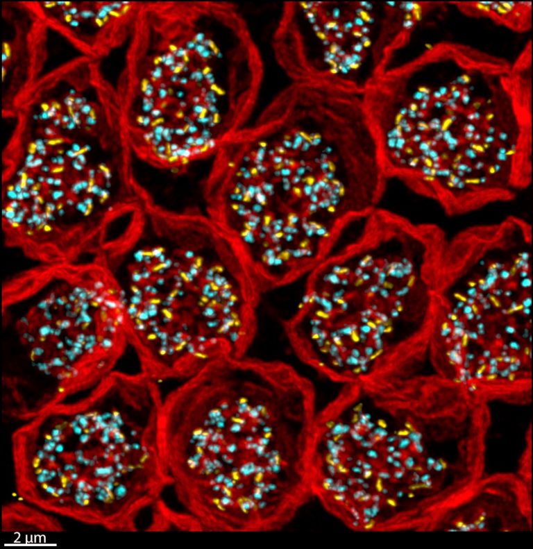
Array of cone photoreceptor terminals labeled for PSD95 (red), the glutamate receptors belonging to one type of bipolar cell (yellow), and glutamate re-uptake transporters (cyan). Courtesy of Steven DeVries, MD, PhD.