
Corneal Graft Rejection Versus Failure
Failure to differentiate the two could result in an irreversible loss of the graft.
KEY TAKEAWAYS
- Unlike graft rejection, graft failure is not an inflammatory process and, thus, does not respond to steroid therapy.
- Early recognition offers the best chance of restoring graft clarity and avoiding progression to graft failure.
Corneal graft rejection and failure are distinct complications of corneal transplantation; however, they are often confused in clinical practice. The reason: They share the clinical presentation of a cloudy, edematous graft and the symptom of decreased vision. Recognizing the difference fast is critical, as the reversal of graft rejection is contingent on immediate aggressive treatment using steroids. Here, we discuss how to differentiate the two.
GRAFT REJECTION
Corneal graft rejection is an immunologic response against the transplant tissue. Depending on which layer is involved, the rejection can be classified as epithelial, subepithelial, endothelial, or mixed. Endothelial rejection is particularly important due to the potential of irreversible endothelial cell loss. The following are implicated as risks of rejection: donor age (< 10 years of age), major histocompatibility complex compatibility, ABO compatibility, and the practices at eye banks, such as preservation status.
- Symptoms. The patient may experience blurry vision, light sensitivity, and eye pain or discomfort, though 30% of patients may be asymptomatic.1
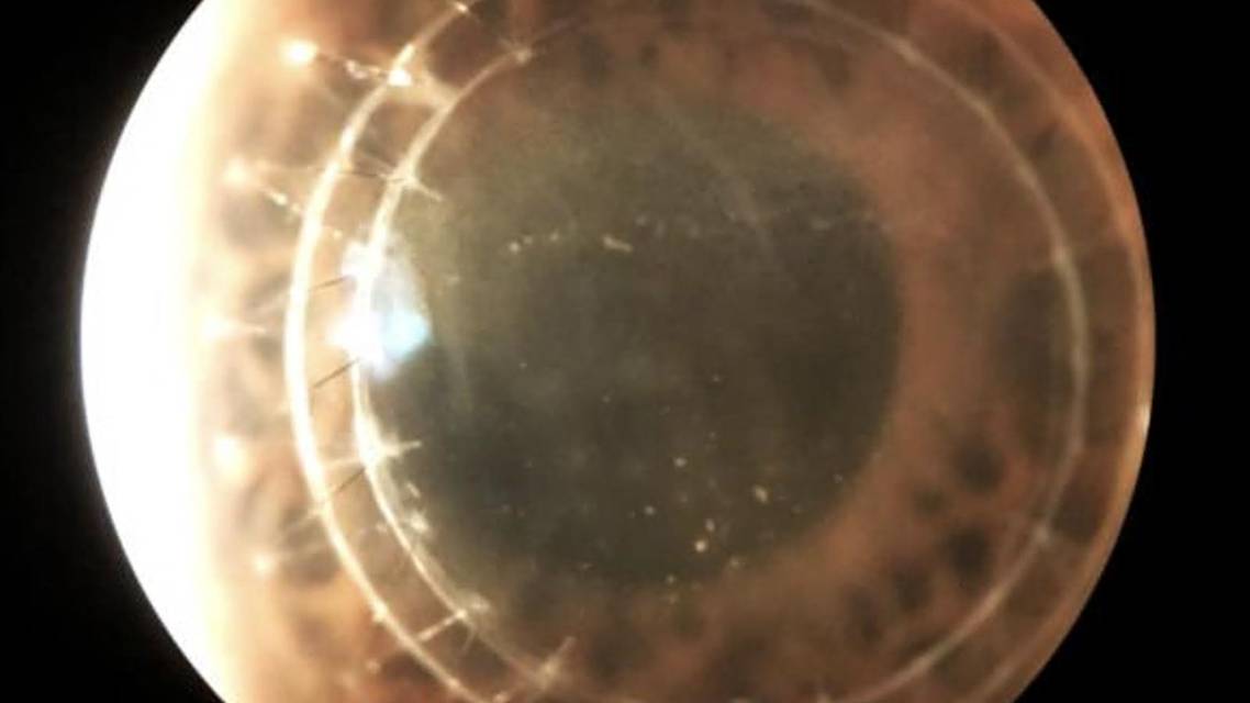
- Signs. Clinically, graft rejection may appear as subepithelial infiltrates, corneal edema, vascularization, a Khodadoust line, and scattered keratic precipitates on the graft itself (Figure).1,2
- Incidence. Although corneal graft rejection has an incidence of ~12% within 2 years, it may still occur several years after transplantation, as rejection remains possible as long as the graft is present.3 The risk of rejection varies by procedure type, with the highest rate occurring after penetrating keratoplasty due to the full-thickness transplantation, followed by Descemet stripping endothelial keratoplasty (DSEK) and Descemet membrane endothelial keratoplasty (DMEK).1 Deep anterior lamellar keratoplasty has a substantially lower risk of rejection, as the recipient’s endothelium is retained.
- Treatment. When rejection is suspected, topical steroids are instilled every 2 hours during waking hours for 1 week, while monitoring the patient closely over the subsequent weeks using visual acuity and pachymetry measurements.2,4 If the corneal edema clears, this supports graft rejection. Once rejection is identified and corneal findings resolve, the steroid regimen is tapered slowly over weeks to months.1,2,4 It is estimated that 50% to 90% of confirmed rejections can be successfully reversed by steroid therapy, allowing recovery of the graft’s clarity and the patient’s visual acuity.2 However, 28% of suspected corneal graft rejections ultimately progress to corneal graft failures.1 The primary reason for this is the irreversible loss of donor corneal endothelial cells during an immune rejection episode.
GRAFT FAILURE
Unlike graft rejection, graft failure is not an inflammatory process and, thus, does not respond to steroid therapy. Additionally, there are two main classifications of corneal graft failure: primary and secondary.
Primary graft failure is caused by the donor cornea having insufficient endothelial function before or at the time of transplantation.
- Symptoms. Patients may report blurred or hazy vision.
Forward-Thinking Outlook
Cultured endothelial cell injection may offer a future alternative to repeat transplantation for selected patients who have endothelial graft failure. A 2026 study in JAMA Ophthalmology of four eyes with failed DSEK grafts reported improved vision and reduced corneal edema after treatment. In addition, investigational endothelial cell therapy AURN001 entered Phase 3 testing in 2026 for corneal endothelial dysfunction. These developments point toward restoring endothelial function, while preserving existing corneal tissue—potentially expanding options for patients facing another transplant.

- Signs. This generally presents as persistent corneal edema in the acute postoperative period, though diffuse stromal and/or epithelial edema can appear on examination due to poor endothelial function.2
- Incidence. This depends on the type of keratoplasty employed. As an example, one study shows the incidence was 1.18% for patients who underwent DMEK and 0.86% for those who underwent DSEK.5
Secondary graft failure usually develops after an initially successful transplant.
- Symptoms. Patients report photophobia, reduced visual acuity, and hazy vision.
- Signs. These include permanent corneal clouding and edema, endothelial cell loss, constant intraocular inflammation, and, sometimes, new blood vessel growth entering the clear donor tissue.
- Incidence. This also depends on the type of keratoplasty. As an example, in the case of penetrating keratoplasty, secondary graft failure was 15.5% by the end of the first year and 40.9% 5 years out.6
- Treatment. Regardless of the classification, treatment for corneal graft failure typically includes repeating the corneal transplant procedure.
THIS OR THAT
A patient who develops corneal edema after transplantation should be examined carefully for corneal graft rejection or failure. Early recognition offers the best chance of restoring graft clarity and avoiding progression to graft failure.
Ready to Claim Your Credits?
You have attempts to pass this post-test. Take your time and review carefully before submitting.
Good luck!
Recommended
 Sept/Oct 2026 ISSUE
Educating Patients on Presbyopia Treatments
Ethin Kiekhafer; Jacob Lang, OD, FAAO, Dipl ABOEthin Kiekhafer; Jacob Lang, OD, FAAO, Dipl ABO